D'Amore Personal Injury Law, LLC
Serious Injury Lawyers Proudly Serving
Baltimore, Annapolis, & Washington, D.C.
Periventricular Leukomalacia
What Is Periventricular Leukomalacia?
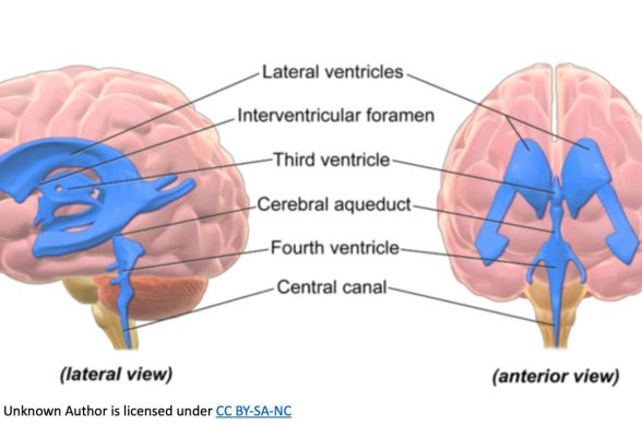
The scientific name for damage and softening of the brain’s white matter is Periventricular Leukomalacia or PVL. The first part, periventricular, refers to the part of the brain around the spaces holding cerebrospinal fluid called “ventricles.” The second part comes from “leuko,” white, and “malacia,” softening.
Periventricular leukomalacia, or PVL, is a brain injury seen in premature babies with very low birth weight. The brain’s white matter is a critical part of the physical structure used to convey signals between the spinal cord and nerve cells and from one part of the brain to another. As the second-most common issue related to the central nervous system in premature deliveries, PVL is a fairly well-known complication for newborn infants.
RELATED ARTICLES
What Causes Periventricular Leukomalacia?
Science doesn’t exactly know the causes that lead to PVL in newborns. Experts believe that a lack of adequate blood supply to the parts of the brain surrounding the ventricles, the fluid-filled areas within the brain, causes the disease. The earlier the baby is born before full-term, the greater the chances of PVL.
The ventricular area of the brain is highly fragile and subject to risks of complications, especially in premature babies whose very delicate brain tissue is just developing. PVL may also be connected to a rupture of the amniotic sac, which is the membrane that holds the fluid that the fetus is in before birth. Other related issues include infection within the uterus, or bleeding inside the brain, called intraventricular hemorrhage.

What Are Some Symptoms Of Periventricular Leukomalacia?
The parts of brain tissue damaged with PVL might cause problems with the nerve cells controlling the child’s motor movements. Damaged nerve cells can lead to muscles that are spastic—excessively tight, which resist movement. Babies born with PVL are at greater risk of cerebral palsy, preventing the child from fully controlling some muscles. The condition can also contribute to learning or developmental challenges.
Diagnostic tests to pinpoint a baby’s developmental issues and to check for PVL might include a cranial ultrasound, using a device much like doctors used to observe the fetus before birth, but focused on the newborn’s brain as measured through the fontanelles. Fontanelles are soft spots where the skull bones meet. Doctors also might order an MRI—magnetic resonance imaging—to assess brain tissue development and further understand the progress of the PVL.
What Should You Expect With Periventricular Leukomalacia?
There is no treatment for PVL, but medical professionals can provide care for the child that may include:
- Physical therapy to work with stimulating and moving muscle groups
- Speech-language therapy to improve communication and understanding
- Vision therapy to improve eyesight
- Occupational therapy as the child grows into an adult to help them find appropriate skills that they can use to function in the world of work
Learn more about what to do after a Brain & Spinal Cord Injury in our FREE guide.
FREE Case Consultation
Fill out the form below and we will contact you.
Or, give us a call at


